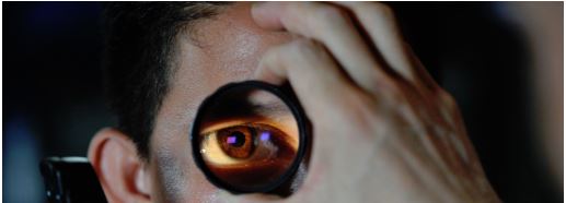
What is an Opthalmascope and What Types are There?
What is an Ophthalmoscope?
An ophthalmoscope is a piece of equipment utilised by ophthalmologists that are used to inspect the internal structure of your eyes, containing the retina. They are used all over the world and are an essential piece of apparatus for all who wish to study the intricate biology of the eye. An expected part of every eye exam, ophthalmoscopes are capable of recognising healthy structures inside the eyeball and clearly help a doctor see indications of ailments in the eye.
The ophthalmoscope itself was invented in 1851 by German physicist Hermann von Helmholtz by using mirrored glass and a conclave lens. It enabled physicians to illuminate the retina and observe it at the same time. This meant that for the first time, they were able to investigate grievances of the eye such as glaucoma, tumours and detached retinas.
Some ophthalmic students may at first think that an ophthalmoscope is extremely similar to a retinoscope, but this is not the case. Both the retinoscope and the ophthalmoscope allow examination of the retina. Although, retinoscopy requires an active light source that may be swiftly moved off the visual axis. The ophthalmoscope is incapable of providing this type of light. On the other hand, the retinoscope is unable to deliver sufficient illumination of the retina to make it beneficial for ophthalmoscopy.
Different types of Ophthalmoscopes
It is important to recognise that not all ophthalmoscopes are handheld devices, including Keeler specialist ophthalmoscopes. An ophthalmologist may find it necessary to use an indirect ophthalmoscope to obtain a wider view of your eye’s interior structure. This is a head visor, worn by the doctor themselves, which cast a bright light, allowing them to better see and enlarge the interior of an eye with the aid of numerous lenses. Read a comparison of the two different types of ophthalmoscopes below.
Direct Ophthalmoscope
A direct ophthalmoscope is the handheld tool that you see in the image on the left of this paragraph. It is an important tool used by all ophthalmologists that constructs unreversed images of around 15 times magnification to examine the rear of the inner eyeball, called a Fundus. Doctors using a direct ophthalmoscope typically carry out the procedure in a darkened room to best see the eyeball.
Physicians use the instrument to examine for variations in the pigment of colour in the interior eyeball as well to see if there is an alteration in the character and calibre of the retinal blood vessels. They will be looking for abnormalities in the macula lutea. This is the part of the retina that receives and examines light from the centre of the visual field. Opacities and macular degeneration of the lens can also be detected through direct ophthalmoscopy.
Indirect Ophthalmoscope
Indirect ophthalmoscopes have demonstrated to be a remarkably respected instrument for the treatment and analysis of detachments, holes, and retinal tears. In order for the acceptable use of an indirect ophthalmoscope, the patient's pupils must be entirely dilated.
What one should you buy?
When deciding what type of ophthalmic instrument you should purchase, you should deliberate about the large amount of patients you'll be treating and the level of diagnosis you'll need to achieve with each of them individually. There will be patients who are coming in for just a simple check-up, which means you may only require the simple view of a direct ophthalmoscope. On the other hand, when you are treating patients that have more serious conditions, the binocular indirect ophthalmoscope could help you identify problems easier and more accurately and prescribe better care for their condition. Take all of this in to account when deciding which ophthalmoscope you should be buying, unless you are required to purchase a certain one at your school or university if studying ophthalmology as they may need you to purchase a certain type or model in order to learn most effectively what they are teaching.


